SLAC topics

Cryo-EM RSS feed

Cryo-EM allows scientists to make detailed 3D images of DNA, RNA, proteins, viruses, cells and the tiny molecular machines within the cell, revealing how they change shape and interact in complex ways while carrying out life’s functions.

Related links:   
Joint institutes and centers  
Cryo-EM fact sheet (pdf) 
Stanford-SLAC Cryo-Electron Microscopy website

Research associate Megan Mayer and graduate student Patrick Mitchell load a sample into a cryogenic electron microscope at SLAC.

News Feature

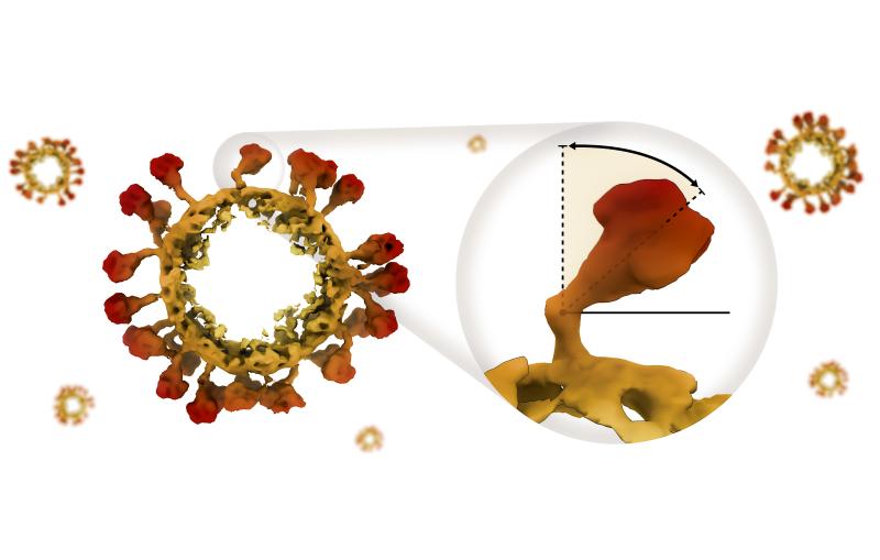
Disabling those hinges could be a good strategy for designing vaccines and treatments against a broad range of coronavirus infections.

A 3D image of a round, spiky coronavirus with inset showing how far its spikes can bend.
News Feature

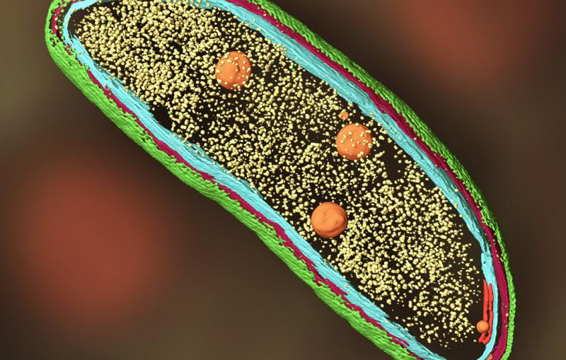
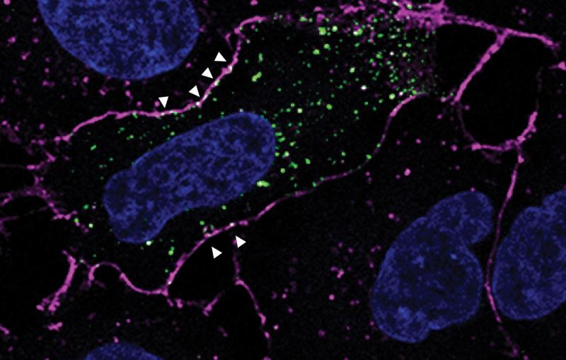
Peter Dahlberg has combined two complex imaging techniques into one. The 2021 Panofsky Fellow adds cryo-ET and biosensors to fluorescence microscopy to give context...

A green, red and blue outline encloses small yellow dots and orange circles, representing parts of a cell.
Press Release

New SLAC-Stanford Battery Center bridges the gaps between discovering, manufacturing and deploying innovative energy storage solutions. 

Illustration showing a battery researcher at left, a battery at center and a grid of battery applications at right.
Illustration

The new SLAC-Stanford Battery Center aims to bridge the gaps between discovering, manufacturing and deploying innovative energy storage solutions. 
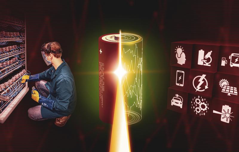
Illustration showing a battery researcher at left, a battery at center and a grid of battery applications at right.
Video

Cryogenic electron microscopy (cryo-EM) is a revolutionary tool for studying the molecular architecture of protein, viruses, cells and the specialized molecular machines within cells...

Stillframe of Cryo-EM explainer video
Video
Past Event

Presented by Peter  D. Dahlberg. Viewing cellular machinery at the nanoscale. 

cryo-EM image of Caulobacter bacterium
Video

Public lecture presented by Peter  D. Dahlberg

cryo-EM image of Caulobacter bacterium
Video
Public Lecture Poster

Viewing cellular machinery at the nanoscale

poster for lecture titled What the cell is going on?
Press Release

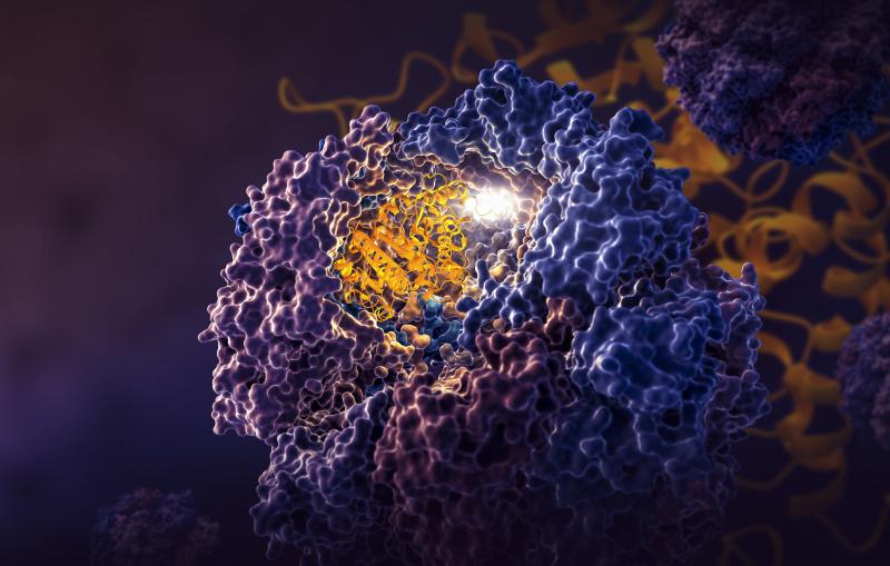
Researchers used cryo-EM (left) to discover how a chamber in human cells (right) directs protein folding. 

A pom-pom like object with curly tangles in purple and blue shades and yellow tangles at center, reminiscent of a zinnia blossom.
Photograph
Lydia-Marie Joubert is pointing at the result of laminating an organic sample down to 100-300nm thickness for cryo-EM imaging. For...
Cryo-EM computer image
Photograph

A scientist works on Tuberculosis samples at the Cryo-EM facility.

A scientist works on Tuberculosis samples at the Cryo-EM facility.
Photograph

Research associate Megan Mayer and graduate student Patrick Mitchell load a sample into a cryogenic electron microscope at SLAC.

Research associate Megan Mayer and graduate student Patrick Mitchell load a sample into a cryogenic electron microscope at SLAC.