
Video
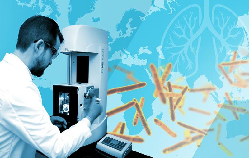
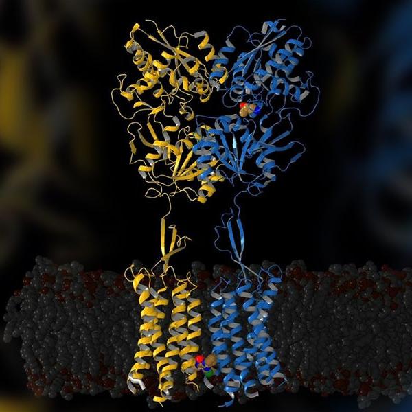
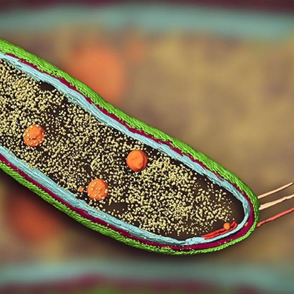
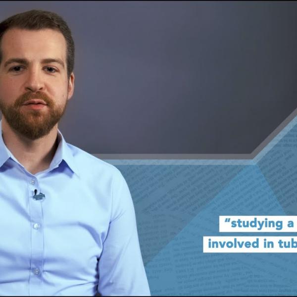
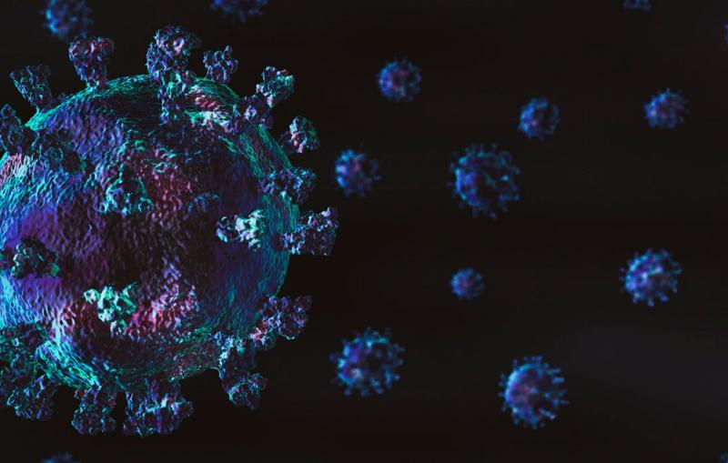
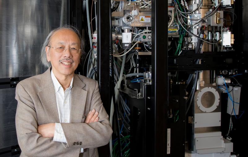
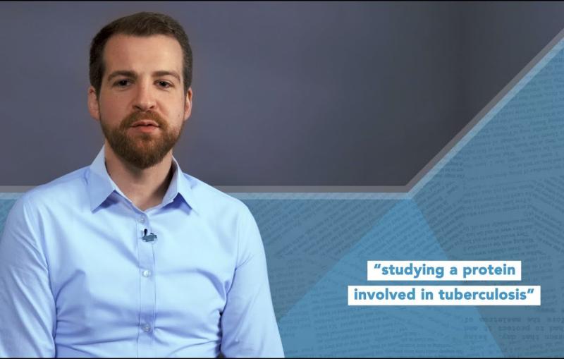
Cornelius Gati and other researchers were studying a protein thought to be important for the progression of tuberculosis when they made a strange discovery...
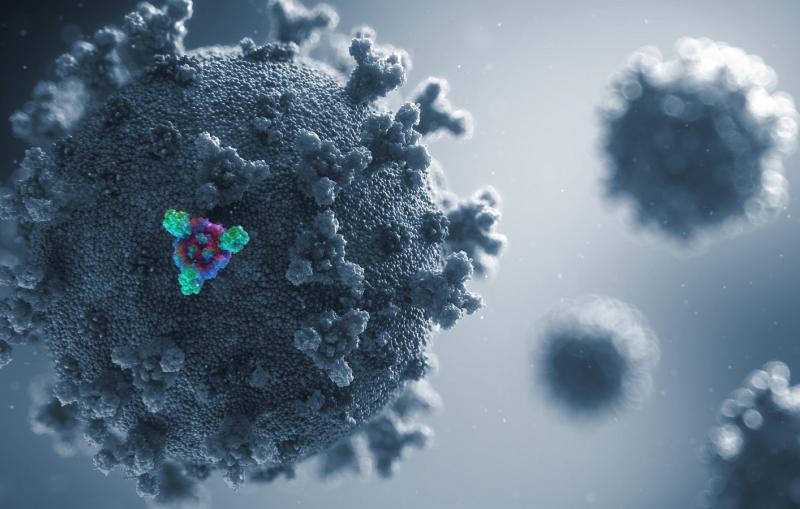
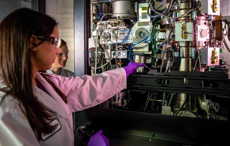
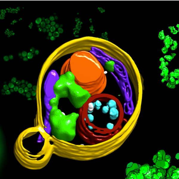
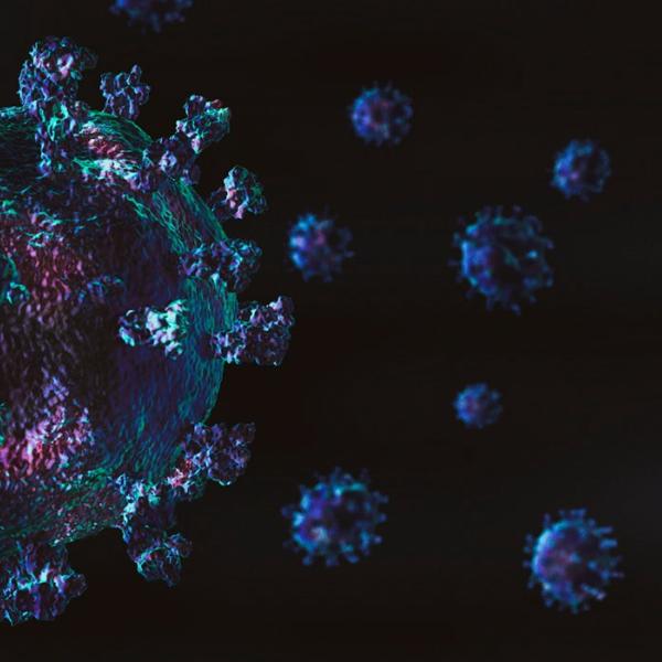
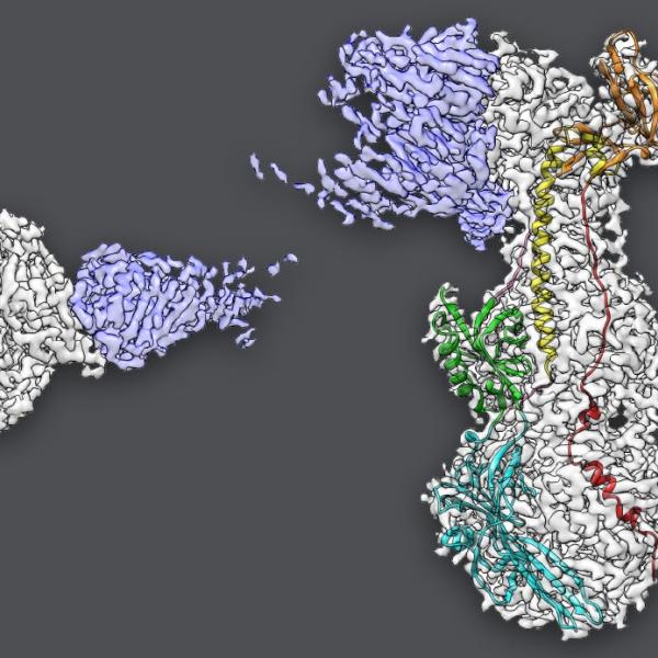
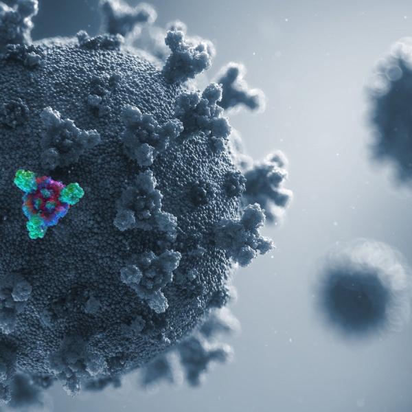
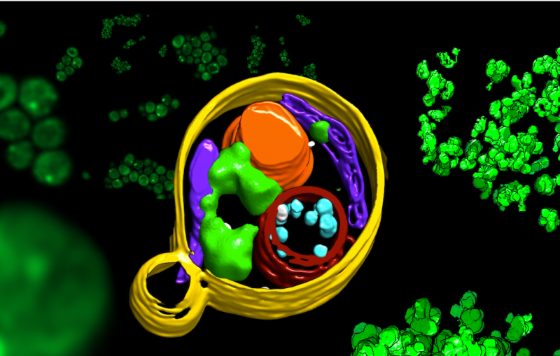
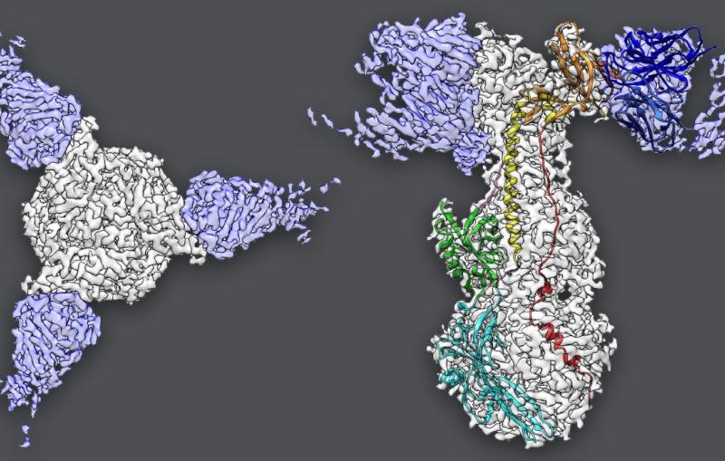
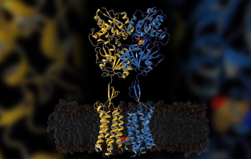
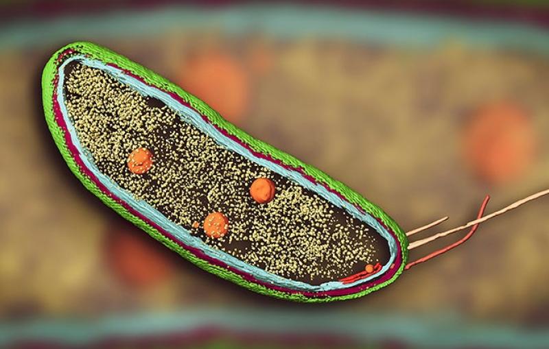
Cryo-EM allows scientists to make detailed 3D images of DNA, RNA, proteins, viruses, cells and the tiny molecular machines within the cell, revealing how they change shape and interact in complex ways while carrying out life’s functions.
Related links:
Joint institutes and centers
Cryo-EM fact sheet (pdf)
Stanford-SLAC Cryo-Electron Microscopy website