High-Speed Movie Aids Scientists Who Design Glowing Molecules
With SLAC’s X-ray laser, a research team captured ultrafast changes in fluorescent proteins between “dark” and “light” states. The insights allowed the scientists to design improved markers for biological imaging.
By Amanda Solliday
The crystal jellyfish swims off the coast of the Pacific Northwest and can illuminate the waters when disturbed. That glow comes from proteins that absorb energy and then release it as bright flashes.
To track many of life’s activities, biologists took a cue from this same jellyfish.
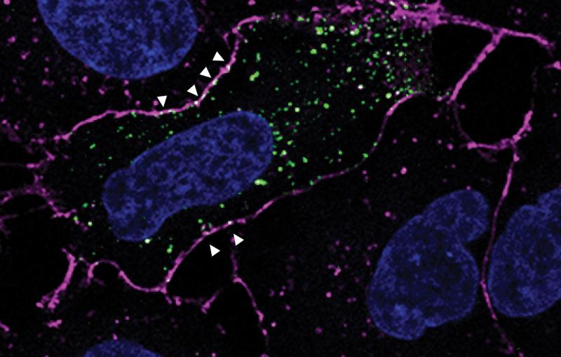
Scientists collected one of the proteins found in the sea creatures, green fluorescent protein (GFP), and engineered a molecular light switch that would glow or remain dark depending on specific experimental conditions. The glowing labels are attached to molecules in living cells so researchers can highlight them during imaging experiments. They use these fluorescent markers to understand how a cell responds to changes in its environment, identify which molecules interact within a cell and track the effects of genetic mutations.
Researchers have studied GFP and other fluorescent proteins for decades to better understand their glowing action and improve their function in scientific studies, but they have never been able to observe the ultrafast changes that occur between “off” and “on” states until now.
In a recent experiment conducted at the Department of Energy’s SLAC National Accelerator Laboratory, a research team used bright, ultrafast X-ray pulses from SLAC’s X-ray free-electron laser to create a high-speed movie of a fluorescent protein in action. With that information, the scientists began to design a marker that switches more easily, a quality that can improve resolution during biological imaging.
“We think that this approach will open a world of possibilities to tailor fluorescent proteins,” says Martin Weik, scientist at the Institute of Structural Biology in Grenoble, France and one of the authors on the publication. “We not only have the structure of the fluorescent protein, but now we can see what is happening between one static state and the other.”
Nature Chemistry published the study on Sept. 11.

Filming a Molecular Light Switch
To observe these intermediate states, the scientists initiated a photochemical reaction in the fluorescent protein with an optical laser at the Coherent X-ray Imaging instrument at the Linac Coherent Light Source, followed by X-ray snapshots at distinct time delays. The optical laser provides energy in the form of photons, mimicking what happens in nature.
“Atoms move around in the photoactive site of the molecule as a result of absorption of a photon,” says Sebastien Boutet, SLAC scientist and a co-author of the paper. “This structural change turns the protein from a dark state to a light-emitting (fluorescent) state.”
There’s a vast body of literature calculating what might happen between the two states, but no one studying the protein was able to see the structural changes in the switch as the photon is absorbed. The molecular switch was just too fast for traditional X-ray imaging techniques.
In this study, the femtosecond X-ray pulses generated by LCLS—arriving in just millionths of a billionth of a second—allowed the team to create stop-action images of the process at an extremely close interval after the proteins were activated by the optical laser.

A Door Half Open
The high-speed snapshots were used to generate a movie starting from the dark state, and gave the researchers insights that they used to design more efficient switchable light-emitting proteins. They found a clue in the time the molecules spent between fluorescent and non-fluorescent states.
“After a picosecond, and for a very short time, this molecular switch is stuck between on and off,” says Ilme Schlichting, scientist at the Max-Planck Institute in Heidelberg, Germany and one of the authors on the publication. “People have predicted this, but to actually visualize its structure is extremely exciting.”
“It’s as if there’s a door and it’s neither closed nor completely open; it’s half open,” she says. “And now we are learning what can go through the door, what might be blocking it and how it works in real time.”
In this study, the scientists found that an amino acid blocked the door and prevented the switch from flipping as easily as possible.
The researchers shortened the amino acid in a mutated version of the fluorescent protein. This engineered version switched more easily and gave better contrast. These traits will allow scientists to observe cellular activity with greater precision.
“Contrast is essential in imaging. It’s like on a TV screen, where to see the best picture, you want the dark to be extremely dark and the color to be super bright and colorful,” says Jacques-Philippe Colletier, a scientist at the Institute of Structural Biology who contributed to the research.
This new molecular movie featuring the jellyfish-inspired proteins lights the way to capture more of life’s microscopic details. The team will continue to fine-tune the protein for other desired characteristics that make it ideal for “super-resolution microscopy,” a type of light microscopy where scientists are able to see illuminated details of cells not distinguishable with conventional light microscopy methods.
The research collaboration included several French institutions, including the Institute of Structural Biology, University of Lille, University of Paris-Sud and the University of Rennes, as well as Max Planck Institutes in Germany and SLAC.
LCLS is a DOE Office of Science User Facility.
Citation: Coquelle et al., Nature Chemistry, 11 September 2017 (10.1038/nchem.2853)
For questions or comments, contact the SLAC Office of Communications at communications@slac.stanford.edu.
SLAC is a multi-program laboratory exploring frontier questions in photon science, astrophysics, particle physics and accelerator research. Located in Menlo Park, Calif., SLAC is operated by Stanford University for the U.S. Department of Energy's Office of Science.
SLAC National Accelerator Laboratory is supported by the Office of Science of the U.S. Department of Energy. The Office of Science is the single largest supporter of basic research in the physical sciences in the United States, and is working to address some of the most pressing challenges of our time. For more information, please visit science.energy.gov.


